
Services
Eye Exams | Diagnostic Testing | Ophthalmic Surgery | OFA Exams | Service Animals

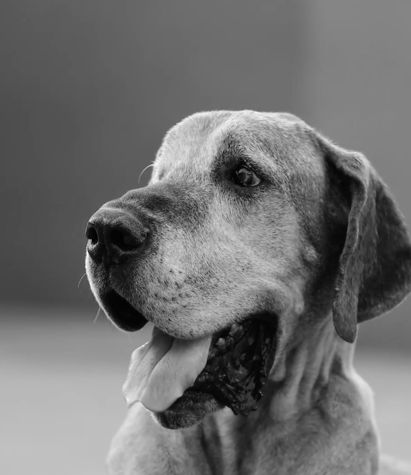
Specialty Eye Exams For All Species
During a comprehensive eye examination, tear production, and intraocular (eye) pressures may be measured and the eyes may be stained with fluorescein stain to assess for corneal injury, tear film quality, and tear duct patency.
Using a slit lamp biomicroscope and indirect ophthalmoscopy, the eyes are evaluated with great magnification to assess for any abnormalities of the eyelids, ocular surface, and interior structures of the eye.


Advanced Diagnostic Testing
Conjunctival Culture
Certain ocular conditions require a sample to be acquired and submitted to a lab for further evaluation. Indications for a conjunctival culture include chronic conjunctivitis, chronic low tear production, and excessive ocular discharge.
Electroretinography
An electroretinogram (ERG) measures the electrical activity of the retina, which is a useful indicator of vision. This test is performed routinely before cataract surgery and is crucial in the diagnosis of certain retinal conditions, such as SARDs.
Fluorescein Stain
Fluorescein stain is used to determine the health of the cornea, tear film, and tear duct. It is especially useful in diagnosing corneal ulcers.
Gonioscopy
Gonioscopy is a procedure performed to exam an eye’s internal drain. It can be helpful in determining the underlying cause of glaucoma, as well as determining if a normal appearing eye is at risk of developing glaucoma.
Histology
Histology is the microscopic study of the surgically removed tissues in and around the eyes of animals. It is used to identify and diagnose various eye diseases and conditions, guiding treatment decisions to preserve comfort and vision.
Maze/Vision Test
A maze test may be used for animals having difficulty seeing. Observing how the pet performs with lights on and lights off, can provide insight on the extent, if any, of vision impairment.
Ocular Ultrasound
Ultrasound allows us to examine the structures in and around the eye in a safe and pain free manner. An ocular ultrasound is routinely performed prior to cataract surgery to ensure the retina is intact behind the opaque cataract. It is also useful for detecting tumors inside of the eye, causes for bleeding within an eye, and lens instability.
Schirmer Tear Test
Normal tear production is essential for the health of an eye. Tear testing allows us to objectively measure the amount of tears produced, in order to diagnose and manage conditions such as dry eye.
Tonometry
Tonometry is a test that measures the pressure within the eye. Knowing the pressure of an eye is critical for conditions such as glaucoma and uveitis.
“There are no words we can use to tell you how thankful we are for Dr Bergstrom. He is an extremely skilled doctor and surgeon. But what sets him apart and makes him truly special is his compassion and the way he cares… not just about his patients, but for all of us parents as well. We were in an unfortunate position to have to make a very quick decision about an eye surgery for our dog Mazie. He clearly laid out the options and the risks and talked through everything with us. But more importantly, he reassured us we were doing the right thing and then hugged us when we cried. We had an amazing outcome…
and are so thankful Dr Bergstrom is in our lives.”
–KELLY
Ophthalmic Surgery
We offer a vast number of traditional and innovative ophthalmic surgeries. Some of the more common procedures are listed below.
Bent Third Eyelid Cartilage Repair
Cherry Eye Repair
Conjunctival Graft for Deep or Ruptured Corneal Ulcers
Conjunctival/Corneal/Eyelid Mass Removal
Cryosurgery
Distichia Hair Removal
Diamond Burr Keratotomy and Bandage Contact Lens Placement
Diode Laser Treatment of Pigmented Iris Lesions
Endolaser Cyclophotocoagulation
Entropion/Ectropion Repair
Enucleation and Orbital Prosthesis
Intracapsular Lens Extraction
Ocular Evisceration and Prosthesis
Phacoemulsification
Retinopexy
Subconjunctival Placement of Sustained Delivery Drug for Dry Eye Disease
Thermokeratoplasty


OFA Exams for Breeders
The Orthopedic Foundation for Animals (OFA) runs the Companion Animal Eye Registry formerly known as CERF. The purpose of OFA eye exams is to provide breeders of dogs with information about genetic eye diseases in order to produce the healthiest possible dogs. Yearly examinations are recommended. Only board-certified veterinary ophthalmologists like Dr. Ben Bergstrom can perform OFA eye exams.
An OFA eye exam is a screening exam. If painful or vision threatening abnormalities are identified during the exam, Dr. Bergstrom will recommend a comprehensive ocular exam. To schedule your dog(s) for an OFA eye exam, please call The Eye Vets at (615) 640-6335. A referral from your primary care veterinarian is not required.
What to Expect
Prior to your dog’s exam, you need to complete the online registration at www.ofa.org/online.
Dr. Bergstrom will enter your dog’s exam results directly into the online form. You will receive an email from OFA with a digital copy of the completed form. After the exam, you can submit your dog’s results to OFA and pay the OFA fees online.
Eye drops will be administered to the dog prior to the examination to dilate the pupils as required by the OFA. Dilation of the pupils will last for several hours but is not painful for the dog. Access to shade should be provided after the exam until the pupils are no longer dilated. Dr. Bergstrom will perform the OFA eye exam approximately 20 minutes after the dilating drops have been administered, and will examine both the front and the back of the eye. The examination itself takes approximately 5 minutes.
Service Animal Eye Exam Event
The Eye Vets is thrilled to offer National Service Animal Eye Exam event every May.
This is a philanthropic event that provides free, ocular screening eye exams to qualified Service & Working Animals each May. These exams are provided by those members of the American College of Veterinary Ophthalmologists who choose to participate and volunteer their time and resources. The program benefits formally trained working animals who selflessly care for us all. Registration is open April 1st - 30th annually. Approximately 8,000 eye exams are provided across America, Canada and Puerto Rico each May.
Registration for the 2025 Event is now open! More information regarding the 2025 Event has been posted to the ACVO website and will continued to be posted on our social media platforms.
For more information please visit: www.acvoeyeexam.org

